. 2022 Jan 14;4(1):e210063. doi: 10.1148/rycan.210063

Figure 4:

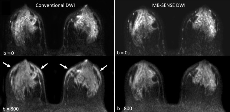
Example of differences in fat suppression observed on images obtained at MB SENSE diffusion-weighted imaging (DWI) compared with those obtained at conventional DWI. On the conventional DWI acquisition (left), unsuppressed fat signal is apparent in both anterior breasts, particularly at b = 800 sec/mm2 (arrows), which was not observed on the MB SENSE DWI acquisition (right), on which tissue contrast is higher. Two readers preferred the image quality of the MB SENSE DWI acquisition, whereas one reader rated them equally. MB = multiband, SENSE = sensitivity encoding.

Example of differences in fat suppression observed on images obtained at MB SENSE diffusion-weighted imaging (DWI) compared with those obtained at conventional DWI. On the conventional DWI acquisition (left), unsuppressed fat signal is apparent in both anterior breasts, particularly at b = 800 sec/mm2 (arrows), which was not observed on the MB SENSE DWI acquisition (right), on which tissue contrast is higher. Two readers preferred the image quality of the MB SENSE DWI acquisition, whereas one reader rated them equally. MB = multiband, SENSE = sensitivity encoding.