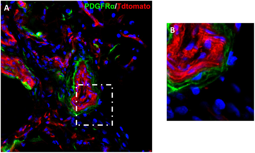
Figure 1. PDGFRα marks a population of cells within the tunica adventitia.
(A) PDGFRαmT/mG reporter mice contain green PDGFRα + cells within the tunica adventitia in the inguinal fat pad . All other cells are red. Nuclear counterstain appears in blue. (B) High magnification of the tunica adventitia.

