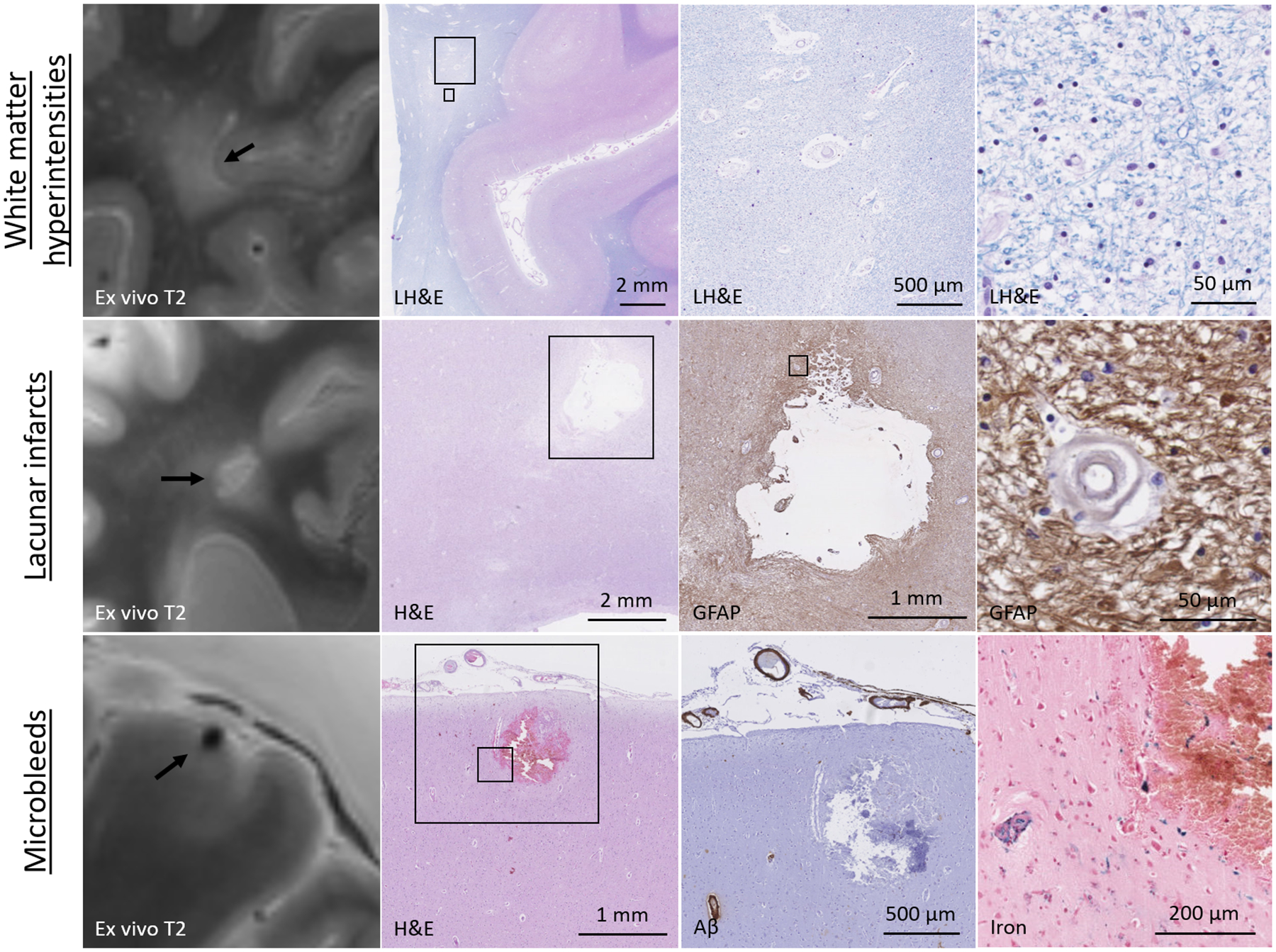
Figure 1.

Neuropathology of white matter hyperintensities, lacunar infarcts, and microbleeds.
Tow row: a representative example of a white matter area with WMH on ex vivo MRI, which corresponded to white matter tissue rarefaction, enlarged perivascular spaces, and demyelination on histopathology. Middle row: a representative example of a lacunar infarct in the white matter on ex vivo MRI, which corresponded to a fluid-filled cavity with a rim of GFAP-positive astrocytes on histopathology. Note the concentric splitting of the wall of a nearby blood vessel. Bottom row: a representative example of a cortical microbleed on ex vivo MRI, which corresponded to a focal area of extravasated red blood cells and iron-positive blood-breakdown products, indicative of a subacute microbleed. Note the presence of vascular amyloid β in nearby blood vessels, but not the vessel responsible for the microhemorrhage.
