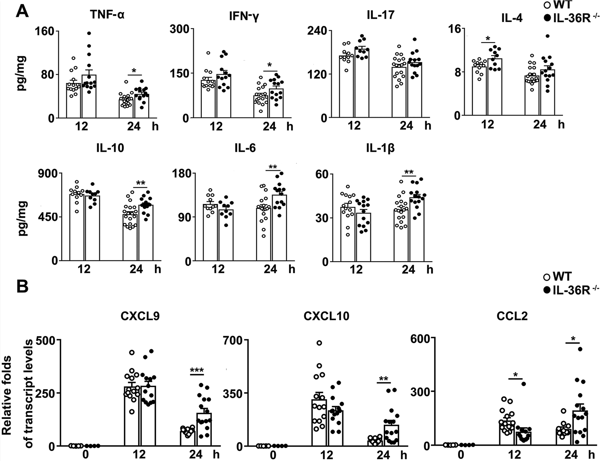
Figure 5. IL-36R-deficiency resulted in increased liver inflammatory responses.

WT and IL-36R−/− mice were i.v. treated with 12 mg/kg of Con A. PBS-injected mice were used as controls (0 h). Protein and RNA were extracted from the livers at 12 and 24 h after Con A treatment. (A) IFN-γ, TNF-α, IL-17, IL-4, IL-10, IL-6 and IL-1β levels in the livers were analyzed by ELISA. (B) Expressions of CXCL9, CXCL10 and CCL2 mRNA in the livers were determined by qRT-PCR. n= 4–5 samples/ control group. n = 10–19 samples/ Con A-treated group from pooled experiments. The data are shown as mean ± SEM of each group from three independent experiments. Two-tailed unpaired T test was used for statistical analysis. * p < 0.05; ** p < 0.01; *** p < 0.001.
