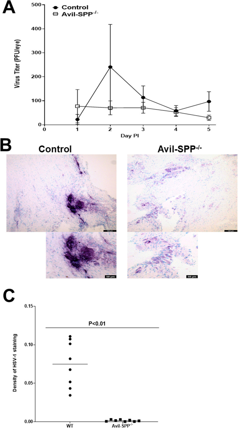
Fig 3. Virus titer in eyes of infected mice and SPP expression in TG of infected mice.

(A) Virus titers in the eyes of Avil-SPP-/- mice. Avil-SPP-/- and control mice were ocularly infected with 2 X 10⁵ PFU/eye of HSV-1 strain McKrae. Tear films were collected on days 1 to 5, and virus titers were determined by standard plaque assay. Each point represents the mean titer of 26 eyes from two separate experiments; (B) HSV-1 protein expression in TG of infected mice. Avil-SPP-/- and control mice were ocularly infected with 2 X 10⁵ PFU/eye of HSV-1 strain McKrae. TG were harvested on day 5 PI and frozen in OCT compound until processing. IHC was performed with anti-HSV-1 antibody and developed using a VectaShield VIP substrate kit. Staining of HSV-1 protein positive cells (purple color) was much stronger in the control group than in the Avil-SPP-/- group; and C) Quantitation of HSV-1 expression density in TG of infected Avil-SPP-/- mice. HSV-1 expression from eight separate IHC staining as (B) above were quantitated by image analysis.
