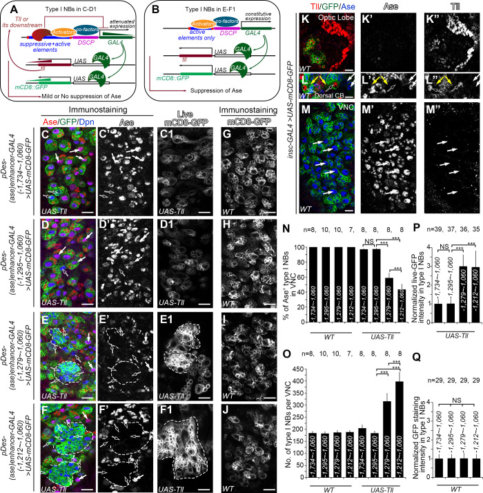
Fig 2. UAS-Tll expression driven by pDes-(ase)enhancer-GAL4 is compromised when the repressive element is included in the GAL4 promoter.
In images (C-F) and (G-J) type I NBs are labeled with mCD8-GFP (in green) driven by pDes-(ase)enhancer-GAL4 drivers containing indicated ase enhancer fragments, and counterstained with anti-Ase (in red) or anti-Dpn (in blue) antibodies. In images (C1-F1), type I NBs are imaged live with the same settings for comparing mCD8-GFP expression levels. In images (L-M) both type I and type II NBs are labeled with mCD8-GFP (in green) driven by insc-GAL4, and counterstained with anti-Tll (in red) or anti-Ase (in blue) antibodies under the same staining condition and imaging settings as those in the image (K). Scale bars equal 10μm. (A-B) Schematic diagrams show how the suppression of Ase by Tll is compromised [A, corresponding to (C-D1)] or maintained [B, corresponding to (E-F1)] in type I NBs. pDes-(ase)enhancer-GAL4 first drives the expression of Tll. The resulted exogenous Tll proteins bind to the repressive element in the pDes-(ase)enhancer-GAL4 driver and attenuates the subsequent GAL4 expression, leading to attenuated mCD8-GFP and Tll expression levels. The attenuated Tll expression is not adequate to suppress Ase expression (A). In contrast, pDes-(ase)enhancer-GAL4 drivers containing only the active elements constitutively drive GAL4 expression, which results in higher expression levels of mCD8-GFP and Tll and subsequent suppression of Ase by Tll (B). (C-D’) Expressing UAS-Tll driven by pDes-(ase)enhancer-GAL4 with the ase enhancer fragment -1,734 ~ -1,060 bps (C-C’) or -1,295 ~ -1,060 bps (D-D’), which contains both the repressive and active elements, leads to the suppression of Ase only in a few type I NBs (open arrows). The majority of type I NBs are still Ase+ (white arrows). (E-F’) Expressing UAS-Tll driven by pDes-(ase)enhancer-GAL4 with the ase enhancer fragment -1,279 ~ -1,060 bps (E-E’) or -1,212 ~ -1,060 bps (F-F’), which contains the active elements only, leads to suppression of Ase in an average of 42% or 57% of type I NBs (open arrows), respectively, and the formation of clusters of supernumerary type I NBs (dashed lines). (C1-F1) mCD8-GFP is expressed at relatively low levels in type I NBs when UAS-Tll is driven by pDes-(ase)enhancer-GAL4 containing the ase enhancer fragment -1,734 ~ -1,060 bps (C1) or -1,295 ~ -1,060 bps (D1), but at much higher levels when UAS-Tll is driven by pDes-(ase)enhancer-GAL4 containing the ase enhancer fragment -1,279 ~ -1,060 bps (E1) or -1,212 ~ -1,060 bps (F1). (G-J) mCD8-GFP is expressed at similar levels in type I NBs when pDes-(ase)enhancer-GAL4 containing the ase enhancer fragment -1,734 ~ -1,060 bps (G), -1,295 ~ -1,060 bps (H), -1,279 ~ -1,060 bps (I) or -1,212 ~ -1,060 bps (J) is used as drivers in wild type VNCs. (K-K”) The robust expression of Tll in optic lobe is detected by anti-Tll antibody (red). (L-L”) Tll expression is detected robustly in type II NBs (open arrows) and weakly in newly born imINPs (yellow arrows), but not in type I NBs (white arrows) in the dorsal central brain (CB). (M-M”) Tll expression is not detected in type I NBs (white arrows) in VNCs. (N-Q) Quantifications of the percentage of Ase+ type I NBs in VNCs (N), the number of total type I NBs per VNC (O), and normalized mCD8-GFP intensity in type I NBs (P, Q) in the VNCs with indicated genotypes. The average mCD8-GFP expression driven by the pDes-(ase)enhancer-GAL4 with the fragment -1,734 ~ -1,060 bps in both the WT and UAS-Tll groups is normalized to 1. Values of the bars are mean ± SD.***, P < 0.001. NS, not significant.

