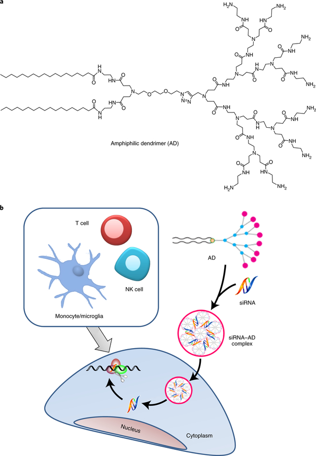
Figure 1:
Schematic presentation of the siRNA delivery mediated by the amphiphilic dendrimer AD. a) The molecular structure of AD. b) Cartoon illustration for the AD-mediated siRNA delivery into various immune cells, including T cells, microglia, and NK cells. (Figures adapted from Refs. 17 and 20). The pink dots represent the amine terminals, while the blue dots represent the amidoamine backbone branching units and the yellow dot represents the triazole ring in AD.

