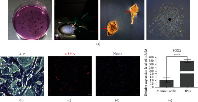
Figure 3.

Preparation and identification of DPCs. (a) An individual whisker HF was isolated, and the DP was separated from isolated HF under stereomicroscopy. Red arrows showed the separated DP condensates. Primary DPCs gradually migrated from the DP condensates after incubation, and DPCs were successfully harvested. (b) ALP staining. Scale bar = 50 μm. (c) Immunofluorescent staining of α-SMA. Scale bar = 100 μm. (d) Immunofluorescent staining of Nestin. Scale bar = 100 μm. (e) Expression of SOX2 in DPCs was detected by qRT-PCR, and rat meniscus cells were used as a control.
