
Figure 4.
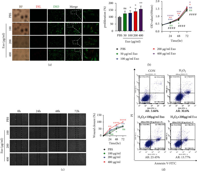
ADSC-Exos were taken up by DPCs and promoted DPC proliferation, migration, and apoptosis inhibition. (a) The internalization of Dil-labeled ADSC-Exos (red) by Dio-labeled DPCs (green) was observed. Bright-field (BF) images were also shown. Scale bar = 200 μm. (b) DPC proliferation was assessed by CCK-8 assay. Data are expressed as the mean ± SD. n ≥ 3; ∗ means P value vs. the PBS group, # means P value vs. the 0 h group. (c) Scratch wound-healing assay. (d) Apoptosis flow cytometry assay. AR: apoptosis rate; Exo: ADSC-Exos. The experiment was repeated three times independently.