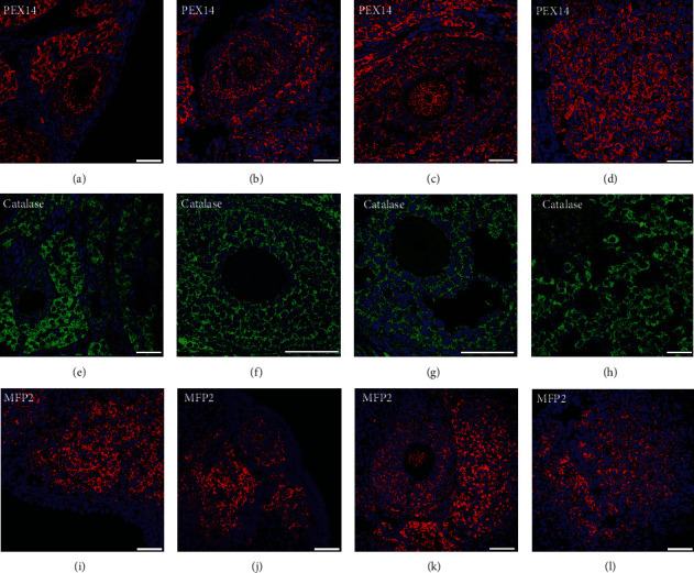
Figure 1.

Immunofluorescence analysis of peroxisomal biogenesis proteins in developing follicles and the corpus luteum. Primary, secondary, tertiary follicles, and corpus luteus were stained with PEX14, catalase, and MFP2 antibodies and with DAPI for cell nuclei. Primary follicles are presented in (a, e, i). Secondary follicles are presented in (b, f, j). Tertiary follicles are shown in (c, g, j), and the corpus luteum is presented in (d, h, j). Scale bars = 40 μm.
