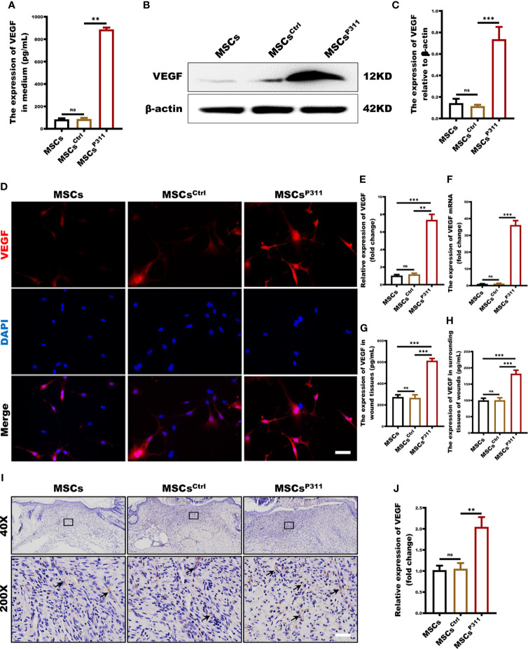
Figure 4.
P311 up-regulates pro-angiogenesis cytokine VEGF secretion of MSCs. (A) The extracellular secretion of VEGF in MSCs, MSCsCtrl and MSCsP311 was examined by ELISA assays. (B–G) The regulation of P311 on VEGF expression of MSCs was estimated by identifying the extracellular secretion, intracellular expression, and mRNA transcription levels of VEGF in MSCs, MSCsCtrl and MSCsP311. (B–E) The intracellular expression of VEGF in MSCs, MSCsCtrl and MSCsP311 was detected by Western blot and confocal microscopy. (B, C) Representative immunoblotting images for VEGF in MSCs, MSCsCtrl and MSCsP311 were shown (left panel), and the gray value of VEGF was statistically analysed (right panel). (D, E) Representative confocal images for VEGF in MSCs, MSCsCtrl and MSCsP311 were shown (left panel), and the MFI value of VEGF was statistically analysed (right panel). Scale bar: 100 µm. (F) The mRNA transcription levels of VEGF in MSCs, MSCsCtrl and MSCsP311 were identified by qRT-PCR. (G–J) The expression of pro-angiogenesis cytokine in wound tissue post-injury was evaluated by assessing the content of VEGF in mice with MSCs, MSCsCtrl, MSCsP311 treatment (n=3/group). Data are representative of at least three independent experiments and represent mean ± SD of indicated number of mice per group. (ns, no statistical significance; **P < 0.01; ***P < 0.001).

