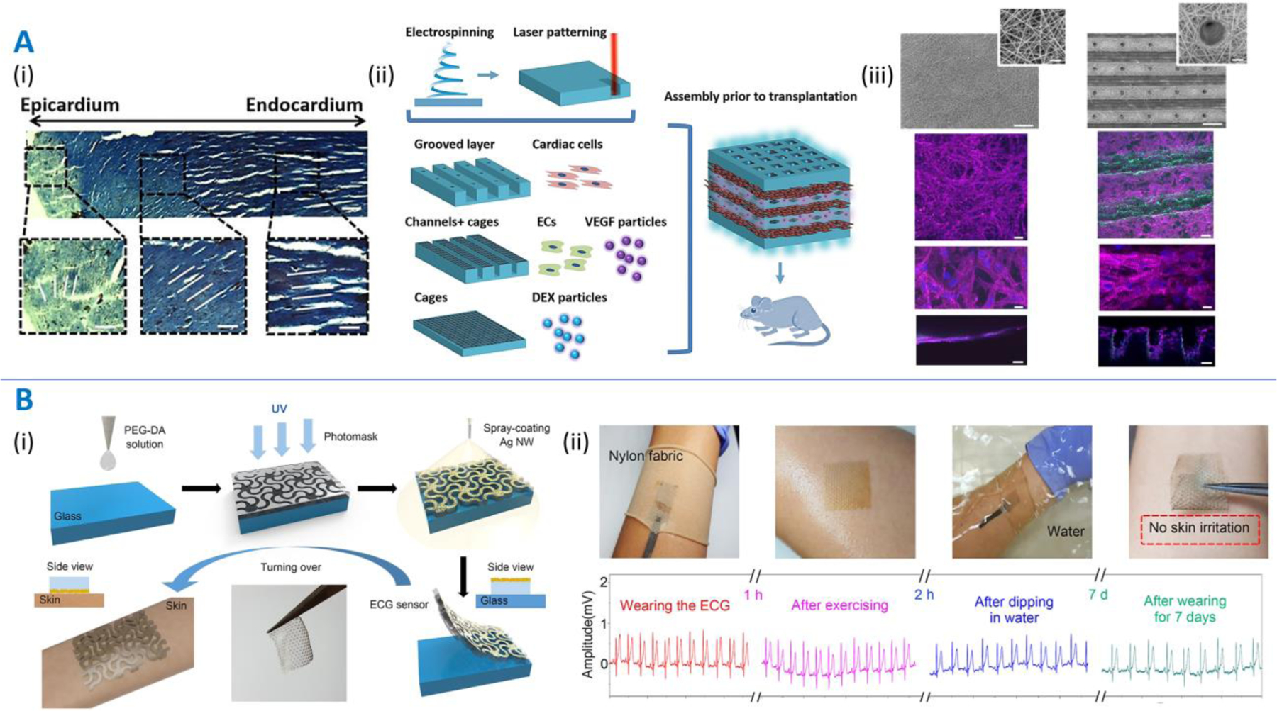
Figure 5. Patches featuring anisotropic (panel A) or auxetic architectures (panel B).

A. Directionally and spatially anisotropic patches. Multi-layered patches mimicking the directional and spatial anisotropy of the human myocardium as demonstrated by Fleischer and colleagues [47]. (i) Masson’s trichrome staining of a transmural section extracted out of the ventricular wall showing variation in fiber orientation in the cardiac tissue. (ii) Scheme of utilizing a bottom-up approach to assembling biomimetic drug (VEGF and dexamethasone)-eluting cardiac patches. (iii) Scanning electron micrographs (top images) demonstrating planar electrospun patches and grooved electrospun patches with micro-holes. Scale bar is 200 µm. Corresponding fluorescence images (bottom) depicting α-sarcomeric actinin (pink) and cell nuclei (blue). Side views are shown in the bottom panels. Scale bars are 50 µm. B. (i) Schematic of the fabrication of auxetic electrode patches developed by Kim and colleagues [73]. PEGDA is dispensed over a glass plate followed by UV crosslinking through a mask and spray coating with Ag nanowires (NWs). (ii) The electrode patches can act as ECG sensors held to the skin via a nylon band, which report changes in ECG measurements depending on the conditions (exercising, in water, etc.) and do not cause any skin irritation over a week. Image reproduced with permission from [47] and [73].
