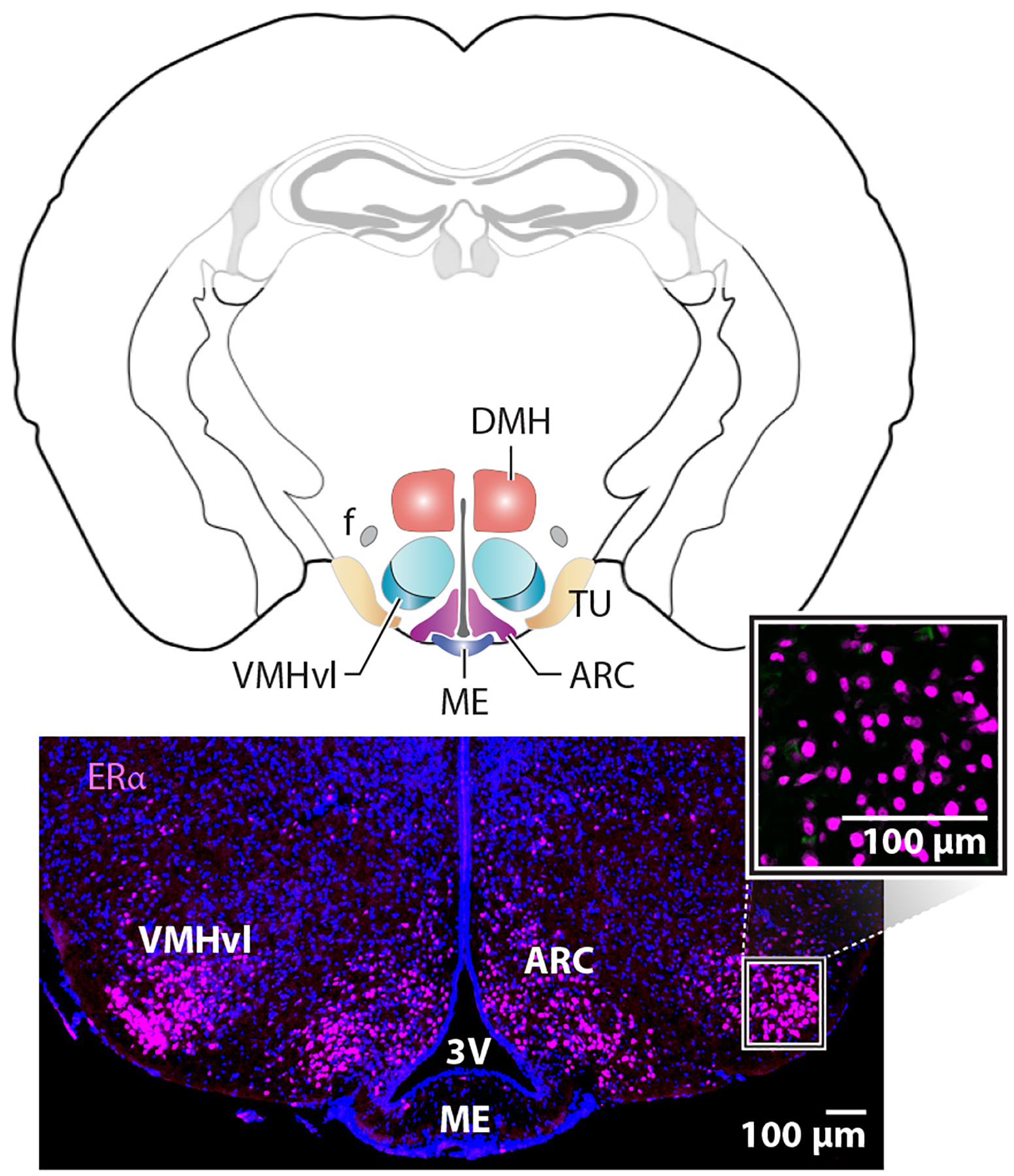
Figure 2.

Enriched expression of estrogen receptor alpha (ERα) in the female medial basal hypothalamus is independent of gonadal hormones. Coronal mouse brain schematic with colored regions of interest, including the ventral lateral region of the ventromedial hypothalamus (VMHvl), the arcuate nucleus (ARC), and the tuberal nucleus (TU). Other landmarks include the fornix (f), dorsal medial hypothalamus (DMH), median eminence (ME), and third ventricle (3V). Lower panels show the expression of ERα in the VMHvl and ARC. The higher magnification panel shows nuclear staining at basal hormone levels or in estrogen-depleted ovariectomized females.
