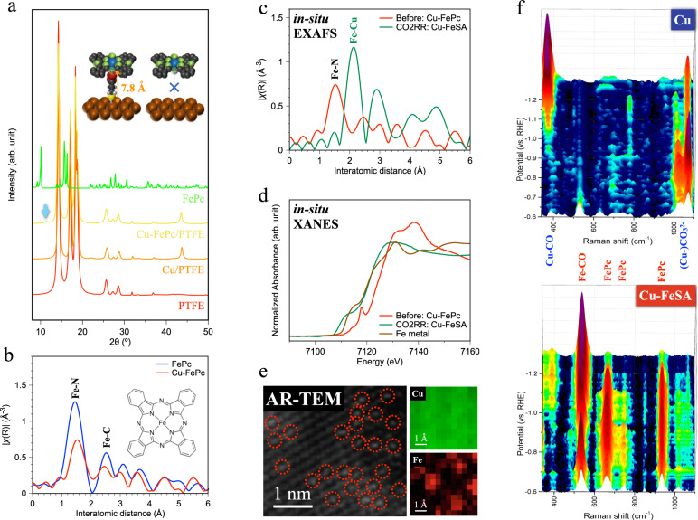
Fig. 3. Materials characterization and in-situ investigation of iron-phthalocyanine-modified and iron-single-atom-anchored copper.
a X-ray diffraction. The inset illustrates the bonding between Cu surface and iron phthalocyanine using 3-mercaptopropionic acid. b Extended X-ray absorption fine structure (EXAFS) of Fe K-edge for the Cu-FePc GDE. c In-situ EXAFS and (d) in-situ XANES of Fe K-edge for identifying Cu-FeSA during CO2RR. e Atomic resolution transmission electron microscope images and atomic elemental mapping using EELS. Dashed circles indicate the single-atom iron. f In-situ Raman spectroscopy for pristine Cu and Cu-FeSA. The intensity scale is 4000 c.p.s. in the spectrum.

