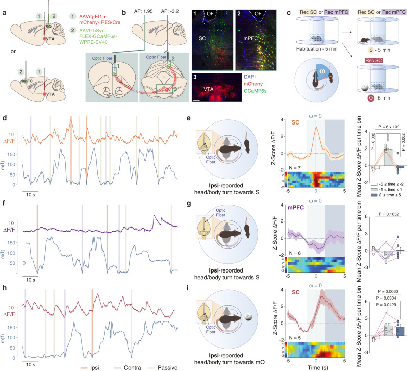
Fig. 2. Calcium activity of SC to VTA-projecting neurons during social and non-social orientation tests.
a Schema of injections of AAVrg-Ef1α-mCherry-IRES-Cre in the VTA and AAV9-hSyn-FLEX-GCaMP6s-WPRE-SV40 in the SC or mPFC. Representative coronal images of midbrain slices of adult mice infected with AAVrg-Ef1α-mCherry-IRES-Cre in the VTA (b panel 3, scale bar: 100 µm) and AAV9-hSyn-FLEX-GCaMP6s-WPRE-SV40 in the SC (b panel 1, scale bar: 50 µm) and in the mPFC (b panel 2, scale bar: 50 µm). The location of the optic fiber (OF) is indicated. Similar viral expression and OF location were observed in all the mice that performed the experiment described in Figs. 2c, 4b. c Schema of the social and non-social orientation test. Bottom left panel: Schema representing the points and vectors used for the calculation of the oriented angle towards the stimulus (ω). d, f, h Example traces. ΔF/F signals recorded in SC- or mPFC-VTA-projecting neurons aligned with instantaneous head orientation ω(t) during orientation test. Ipsi-recorded and contra-recorded head/body turn episodes are reported as well as the passive crossing events. e Left panel: Schema of the ipsi-recorded head/body turns towards the social stimulus (S). Middle panel: Peri-event time histogram (PETH) of normalized ΔF/F for SC-VTA-projecting neurons, centered on ipsi-recorded orientation towards social stimulus. Right panel: Mean ΔF/F (Z-score) before, during and after ipsi-recorded orientation. Repeated measures (RM) one-way ANOVA (Events main effect: F(2,6) = 44.34, P < 0.0001) followed by Bonferroni-Holm post-hoc test correction. g Left panel: Schema of the ipsi-recorded head/body turns towards the social stimulus (S). Middle panel: PETH of normalized ΔF/F for mPFC-VTA-projecting neurons, centered on ipsi-recorded orientation towards social stimulus. Right panel: Mean ΔF/F (Z-score) before, during and after ipsi-recorded orientation. RM one-way ANOVA (Events main effect: F(2,5) = 2.17, P = 0.1652). i Left panel: Schema of the ipsi-recorded head/body turns towards the moving object (mO). Middle panel: PETH of normalized ΔF/F for SC-VTA-projecting neurons, centered on ipsi-recorded orientation towards moving object. Right panel: Mean ΔF/F (Z-score) before, during and after ipsi-recorded orientation. RM one-way ANOVA (Events main effect: F(2,4) = 10.34, P = 0.0060) followed by Bonferroni-Holm post-hoc test correction. N indicates the number of mice. All the data are shown as the mean ± s.e.m. as error bars or error bands. Source data are provided as a Source Data file.

