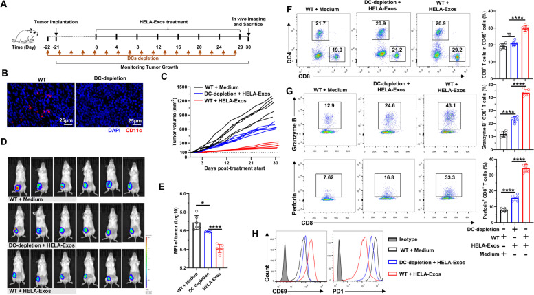
Fig. 6.
DC depletion limited the antitumor efficacy of HELA-Exos in vivo. DC depletion models were established as described in the methods section, and therapy was initiated. The mice were sacrificed at Day 30 after treatment initiation. Tumor tissue immune infiltrates were analyzed by flow cytometry. A Schematic diagram of the vaccine dosing regimen for DC-depleted or nondepleted BALB/c mice with orthotopic breast cancer. B DC depletion efficiency was confirmed by IF. Blue: DAPI; Red: CD11c. Scale bar, 25 μm. C Tumor size was measured every other day after treatment initiation. Tumor growth curves for each mouse are shown. D and E In vivo tumor imaging in mice was performed using an IVIS Spectrum In Vivo Imaging System at Day 30 after initiation of therapy, and the bioluminescence intensity of the tumor was proportional to the size. F The percentages of tumor-infiltrating CD4+ or CD8+ T cells were measured using flow cytometry. G and H Tumor-infiltrating CD8+ T cells were analyzed for the expression of granzyme B, perforin, CD69, and PD1 by flow cytometry. The data are presented as the mean ± SD; n = 6. A t test was performed for statistical analysis (****: P < 0.0001; *: P < 0.05 ns: P > 0.05)

