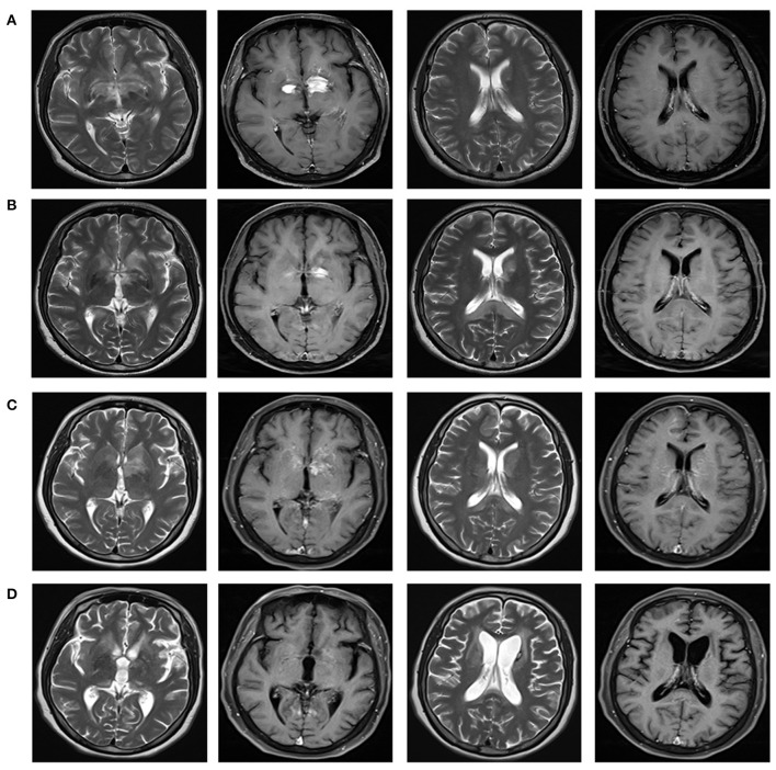
Figure 2.
Brain magnetic resonance imaging reflecting the imaging changes of lesions in bilateral basal ganglia and around the third ventricle, and the changes of splenial corpus callosum lesions during the course of disease. Brain MRI with T2-weighted and T1 contrast-enhanced were shown in different columns. (A) In December 2020, multiple abnormal enhancement lesions in bilateral basal ganglia and around the third ventricle were demonstrated. (B) In January 2021, lesions in bilateral basal ganglia and around the third ventricle reduced significantly with less enhancement. A new T2 signal in the splenium of the corpus callosum without enhancement was shown. (C) In March 2021, lesions in bilateral basal ganglia and around the third ventricle were the same as before, with increased enhancement. Attenuated T2 signal abnormality in the splenium of the corpus callosum was observed. (D) In August 2021, lesions in the bilateral basal ganglia and around the third ventricle significantly reduced, with complete resolution of the enhanced lesions. Abnormal signal foci in the splenium of the corpus callosum disappeared.

