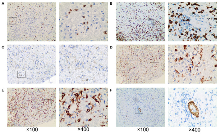
Figure 4.
Pathological examination revealed extensive perivascular inflammation. (A) CD8+ T cells and (B) CD3+ T cells were infiltrated throughout the brain parenchyma, rather than (C) CD4+ T cells. Prominent perivascular cuffing of (D) CD68+ and (E) CD163+ macrophages were seen scattered around small vessels and in the parenchyma. (F) The vessel wall indicated by SMA was intact. ×100 and ×400.

