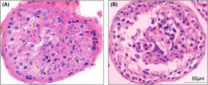
FIGURE 1.

Pathological haematoxylin and eosin staining of testicular tissues from an OA control and the NOA patient. (A) The testicular puncture tissue of an OA control with normal spermatogenesis. The black arrows point to the primary spermatocytes in the pachytene phase, and the yellow arrows point to the mature sperm cells. (B) The testicular tissue of the NOA patient. The red arrows point to the blocked primary spermatocyte without mature forms. White arrowheads indicate Sertoli cells both in OA and NOA samples. NOA, non‐obstructive azoospermia (NOA); OA, obstructive azoospermia
