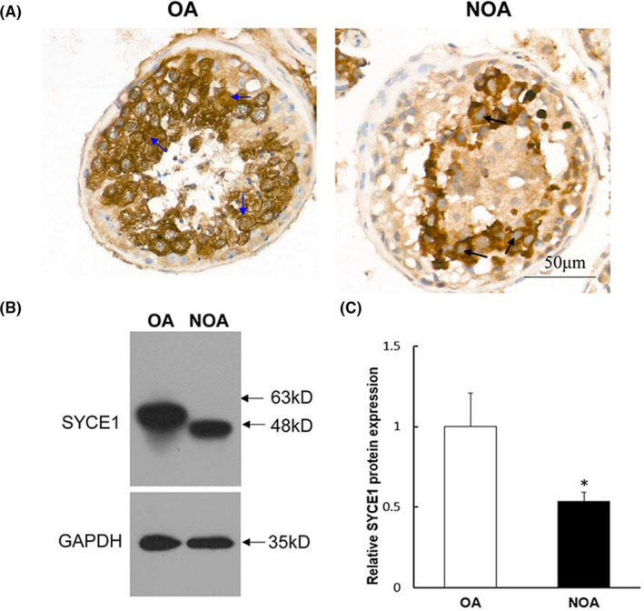
FIGURE 3.

Examination of mutated SYCE1 protein expression and localization in the NOA patient. (A) Immunohistochemical staining showed the endogenous expression and localization of wild‐ and mutated‐type SYCE1. The blue arrows indicated that SYCE1 was expressed in the nucleus of spermatocytes of the OA control. The black arrows indicated that SYCE1 was undetected in the nucleus of arrested spermatocytes of the NOA patient. (B) Western blotting (WB) was used to study the effect of SYCE1 mutation on its endogenous protein expression. The total protein was extracted from the whole blood cells, and GAPDH was selected as the internal control of WB. (C) Grey values (IntDen) of WB bands in (B) were calculated, and the relative protein expression difference between groups was counted. ‘*’ represented statistically significant difference. NOA, non‐obstructive azoospermia; OA, obstructive azoospermia
