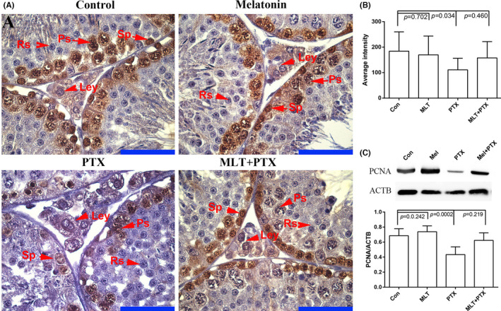
FIGURE 4.

Expression of PCNA in in control and PTX‐treated mice testes Testes were obtained from control, MLT‐treated, PTX‐treated and MLT+PTX‐treated mice. The sections were stained by haematoxylin and eosin. Mel, MLT; PTX, PTX. A, immunostaining of PCNA in testes; B, quantification expression of PCNA in testes; C, Western blot analysis of PCNA in testes in triple replications. The data were analysed through one‐way ANOVA, p < 0.05 was considered as significant. Each bar represents 50 μm
