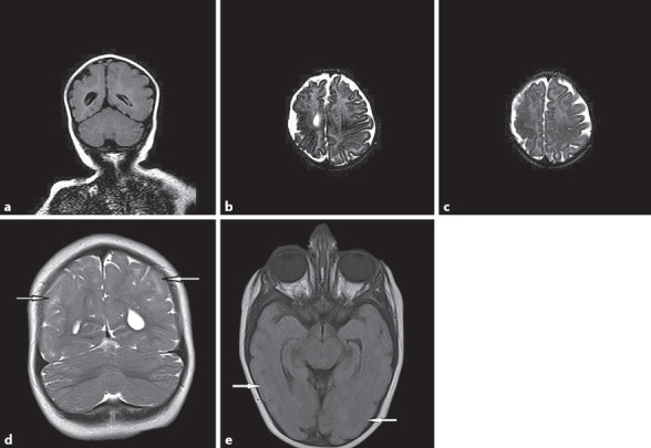
Fig. 2.

Radiological findings. Patient 1: a Symmetrical mild ventriculomegaly, simplified gyral pattern, and cortical dysplasia located in the right temporaparietal lobe on T2 FLAIR coronal image. b, c T2-weighted axial images of brain. Patient 2: d Pachgyria pattern of cortical thickness on coronal T2-weighted image. e Axial FLAIR image.
