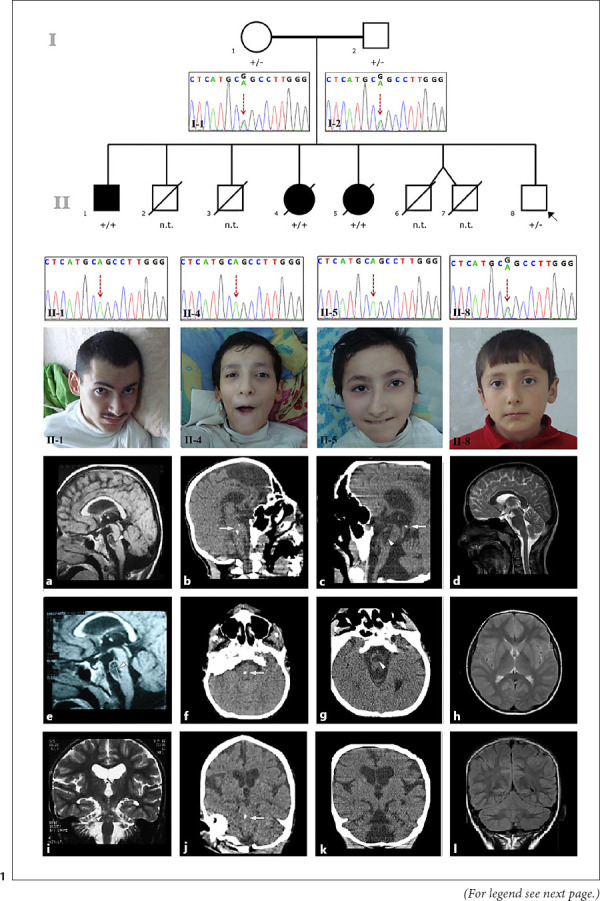
Fig. 1.

Pedigree and electropherograms of the study family and photographs of the affected siblings (brother, II-1; older sister, II-4; and younger sister, II-5) and the proband, II-8. Brain MRI of the brother showed a 20.4 × 12.7 mm intraparenchymal pontine cyst on sagittal T1-weighted and fluid attenuation inversion recovery images (a, e, arrowhead) and cerebellar atrophy on a coronal T2-weighted image at the age of 22 years (i). Cranial CT image of the older sister showed pontine calcification on sagittal (b), axial (f), and coronal (j) images (arrows) at the age of 22 years. Cranial CT image of the younger sister showed a pontine cyst (arrowhead) and calcification on a sagittal image (c, arrow), pontine cyst on an axial image (g, arrowhead), and cerebellar atrophy on a coronal image (k) at the age of 20 years. Brain MRI of the proband showed normal sagittal T2-weighted (d), axial T2-weighted (h), and coronal fluid attenuation inversion recovery (l) images at the age of 10 years. n.t., not tested; +/+, homozygous for the variant; +/−, heterozygous for the variant.
