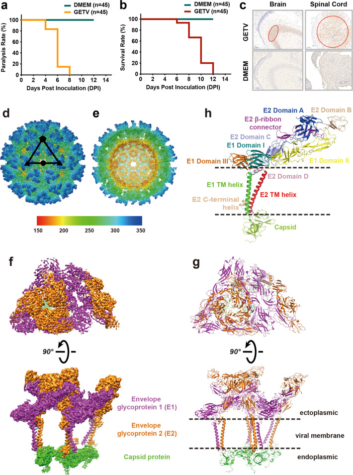
Fig. 1. Overall structure of the infectious GETV virion determined by cryo-EM at 2.8 Å resolution.
a–c Ninety 3-day-old mice were randomly divided into two groups and inoculated oronasally with strain GETV-V1 suspension or an equal amount of DMEM. a Motor disabilities displayed in GETV-inoculated mice at 4 DPI; by 8 DPI, all of the GETV-inoculated mice displayed paralysis. b Mice inoculated with GETV died at 6 DPI, and all of the inoculated mice were dead by 12 DPI. c Tissue from hippocampal dentate gyrus in the brain and spinal cord under lumbar vertebrae were subjected for immuno-histochemistry analysis. There were abundant GETV antigens distributed in the brain and spinal cord under lumbar vertebrae in the inoculated newborn mice (highlighted in red circles). d–h Cryo-EM structure of GETV at 2.8 Å resolution. d GETV virion showing the external surface with assigned symmetry axes. e Central cross-section of the GETV density map. f, g Density map and atomic model of one asymmetric unit. The E1, E2, and capsid proteins are colored separately in magenta, orange and green. h Atomic model of E1–E2–capsid heterotrimer. Different subdomains of E1 (Domain I, II, III, and TM helix) and E2 (Domain A, B, C, D, TM helix, and C-terminal helix) are shown, following the previous definition for alphaviruses33,34.

