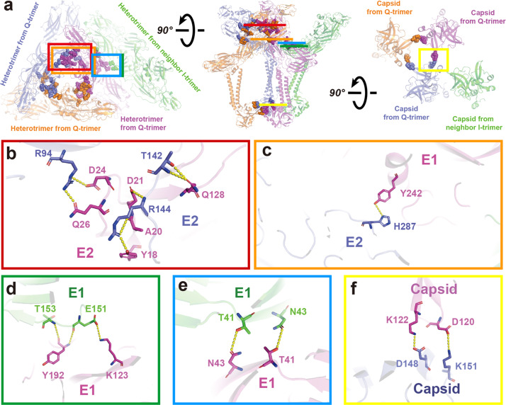
Fig. 3. Interaction of GETV protein domains in the ASU.
a The top, side, and bottom view of the ASU. Four E1–E2–capsid heterotrimers in the ASU are colored slate, magenta, orange, and green. Residues involved in the interaction are displayed as spheres with the same color as the corresponding heterotrimers. The slate, magenta, and orange heterotrimers form the Q-trimer, and the green heterotrimer is from the neighboring I-trimer. b Zoomed-in view from the red box in a of the interaction between E2 (magenta) and the neighboring E2 (slate) protein. c Zoomed-in view from the orange box in a of the interaction between E1 and the neighboring E2 protein. d, e Zoomed-in views from the green and blue boxes in a, showing the interactions between E1 (magenta) and the neighboring E1 (green), which also represent the interactions between Q-trimer and I-trimer. f The interaction between one capsid protein (magenta) and a neighboring capsid protein (slate) in the Q-trimer. The yellow dashed lines indicate the distance between the atoms involved in the interaction (the cut-off distance is 3.5 Å).

