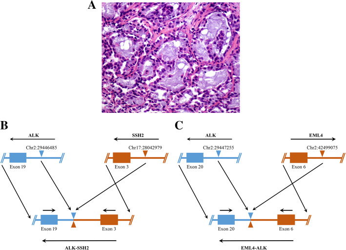
Fig. 2.
Pathological examination and schematic diagram of ALK rearrangement of case 1. (A) Hematoxylin and eosin (H&E) staining of the left upper lobe revealed adenocarcinoma (× 400). (B) A breakpoint within ALK (shown in blue) intron 19 at chromosome 2 was fused within SSH2 (shown in red) intron 2 at chromosome 17, giving rise to the ALK-SSH2 fusion gene. (C) A breakpoint within ALK (shown in blue) intron 19 at chromosome 2 was fused within EML4 (shown in red) intron 6 at chromosome 2, but have opposite orientations, giving rise to the EML4-ALK fusion gene

