Fig. 1.
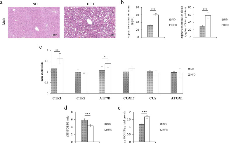
Serum and tissues copper concentrations, redox balance and MG-H1 levels in ND or HFD mice. a Representative photomicrographs of Hematoxylin and Eosin staining in liver sections from representative liver tissues of male mice fed with ND or HFD. Original magnification ×10. b Copper concentration in serum and liver samples of ND and HFD mice. c Transcriptional levels of CTR1, CTR2, ATP7B, COX17, CCS and ATOX1 genes. d Levels of reduced GSH expressed by tGSH/GSSG ratio and d MG-H1 (methyl-glyoxal-hydro-imidazolone) protein adducts. Values are expressed as means ± SD and data were analyzed by a t-Test analysis (*P < 0.05; **P < 0.01; ***P < 0.001)

