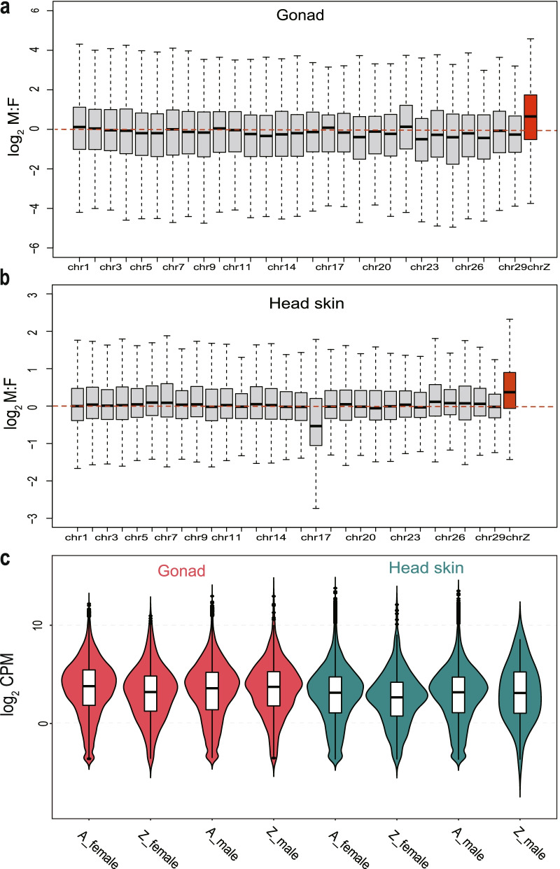
Fig. 2.
Comparison of the expression levels of autosomal-linked and Z chromosome-linked genes (CPM > 0). The M:F (male:female) ratio distributions on the Z chromosome and the autosomes for the gonad (a) and head skin (b). c Violin plots of the Z chromosome and autosome gene expression for each tissue in males (ZZ: AA) and females (Z: AA). Read counts were converted to counts per million (CPM) values to measure the expression of genes. Values are represented on a log2-transformed CPM scale (outliers of |log2 FC| > 5). For simplicity, chromosomes W, 31, and 33 are not included. The red lines indicate equal expression levels between females and males on the chromosome. **** P < 0.01, ns P > 0.05

