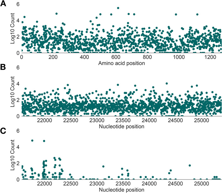
Fig. 1.
Genetic diversity in the spike protein of all available SARS-CoV-2 genome sequences. The vertical axis of each plot shows the number of designated sequences that exhibit mutations at each amino acid position (log10-scaled). A Distribution of non-synonymous variation across amino acid positions of the spike protein (horizontal axis). B Distribution of synonymous variation across nucleotide sites in the spike protein (horizontal axis). C Distribution insertion and deletion variation (indels) across nucleotide sites in the spike protein (horizontal axis). Each point represents the 5′ nucleotide site at the start of each indel. In each plot, genetic variation is determined by comparison with a lineage A reference sequence (Wuhan/WH04/2020, EPI_ISL_406801)

