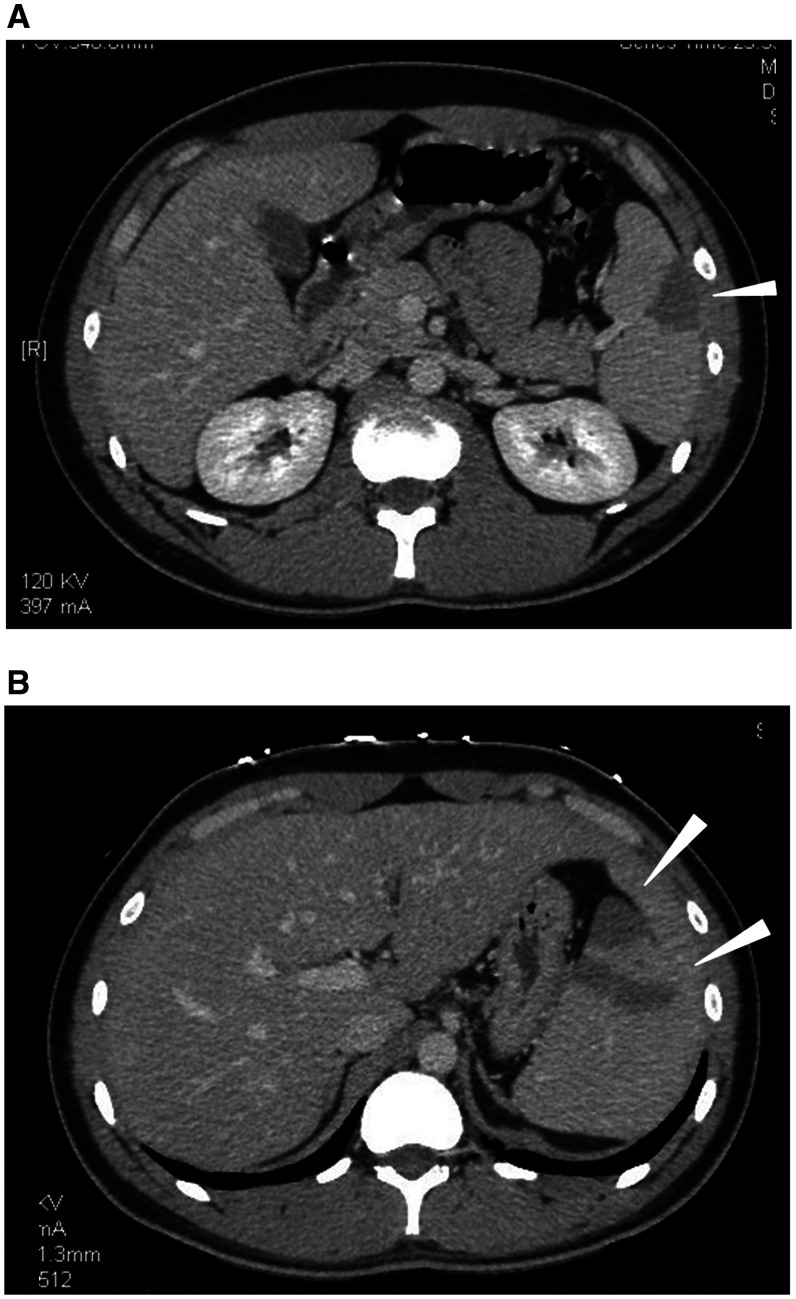
Figure 1.
(A) On day 0, a contrast-enhanced computed tomography (CT) of the abdomen shows wedge-shaped, low-density defect in the peripheral area of the spleen (white arrowhead) in favor of splenic infarction. (B) On day 1, a contrast-enhanced CT of the abdomen reveals new multiple low-density defects in the spleen (white arrowheads).

