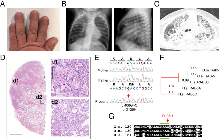
Fig. 1.
Clinical features of the proband with a de novo variant in RAB5B. (A) Shortened broad fingers with nail clubbing. (B) Hyperinflation of lungs and interstitial opacities by chest radiograph (posterior–anterior and lateral views) at 7 mo of age. (C) Computed tomography of the lung at 11 mo of age shows acute and chronic interstitial changes. (D) Lung biopsy stained with hematoxylin and eosin (higher-power details are in insets d1 and d2) shows alveolar filling (d1; asterisk), AT2 cell hyperplasia (d1; bracket), remodeling (d2), and fibrosis (d2). (Scale bars: D, 2.5 mm; d1 and d2, 100 μm.) (E) Dideoxy (Sanger) sequencing traces from parents and the proband showing a de novo heterozygous variant in RAB5B, c.406G > C, p.Asp136His. (F) Phylogenetic tree of Rab5 indicating the evolutionary orthologs and human paralogs. Alignment distance values are shown (Clustal Omega). C.e., C. elegans; D.m., D. melanogaster; H.s., Homo sapiens. (G) Alignment of the RAB5B sequence from H.s. residues 121 to 151 with orthologs from indicated species. The location of the variant edited in C. elegans corresponding to the conserved aspartate [D] in the proband is indicated (red arrow). The fourth region of the conserved nucleotide binding domain (NKXD) is indicated (green bars above and below). Identical residues are shaded black; conserved residues are gray (SI Appendix, Fig. S2).

