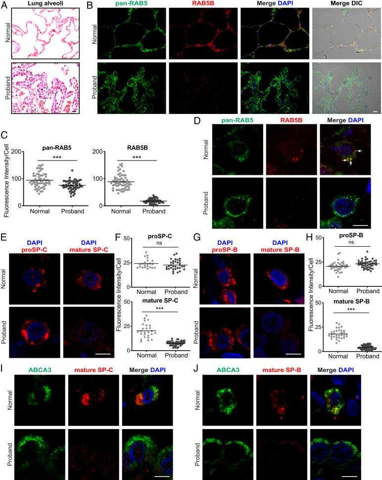
Fig. 4.
Loss of RAB5B and mature SP-B and SP-C in the proband lung. Lung sections from normal donor and proband lung tissue are stained as indicated. (A) AT2 cell hyperplasia in the proband (hematoxylin and eosin). (B) Immunostaining for pan-RAB5 and RAB5B (SI Appendix, Figs. S13 and S14). Differential interference contrast (DIC) microscopy, together with staining, shows lung structure, with a pentagonal alveolar organization in the normal donor and hyperplastic disorganization in the proband. (C) Quantification of immunostaining from B. (D) Confocal microscopy images of single lung cells with pan-RAB5 and RAB5B antibodies showing colocalization in cytoplasmic puncta (arrows). (E) proSP-C and mature SP-C staining and (G) proSP-B and mature SP-B staining in single cells. (Scale bars: A and B, 20 μm; D, E, and G, 5 μm.) (F and H) Quantification of immunostaining from low-magnification staining for proSP-C, mature SP-C, proSP-B, and mature SP-B. (I and J) ABCA3 and mature SP-C staining and ABCA3 and mature SP-B staining, respectively, in individual cells. Images in A, B, D, E, G, I, and J are representative of n = 3 to 4 normal donor lungs. Nuclei were stained with 4′,6-diamidino-2-phenylindole (DAPI). Shown in C, F, and H are plots of relative fluorescent intensity of cells from n = 4 to 6 images from each condition analyzed by the two-tailed Student’s t test. ns, not significant. ***P < 0.001.

