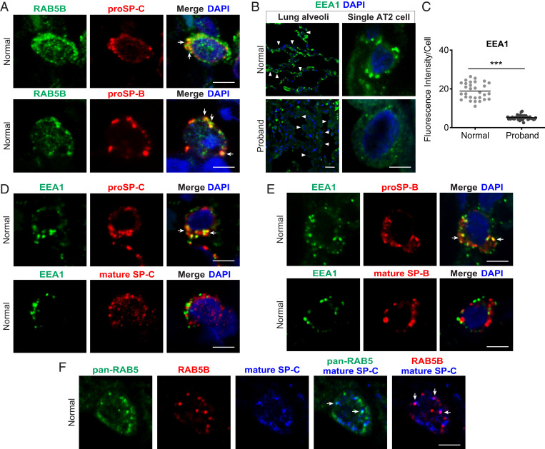
Fig. 5.
Colocalization of proSP-B and proSP-C with RAB5B and EEA1 in donor AT2 cells. Tissue sections from normal donor lung were immunostained with antibodies for the indicated protein. (A) Colocalization of RAB5B and proSP-C or RAB5B and proSP-B (arrows) in single cells. (B) EEA1 staining. (Upper) Normal donor. (Lower) Proband. (Left) Low-power image; arrowheads indicate a subset of AT2 cells. (Right) Single-cell confocal micrographs are shown. (C) Quantification of EEA1 staining from low-power images in B. ***P < 0.001, two-tailed Student’s t test. (D and E) Normal donor lung EEA1 and proSP-C or mature SP-C and EEA1 with proSP-B or mature SP-B, respectively, in single cells. (F) Pan-RAB5, RAB5B, and mature SP-C in normal lung. Arrows in A, D, E, and F indicate colocalization of markers in cytoplasm. Images in A, B, D, E, and F are representative images of n = 3 to 4 normal donor lungs. Nuclei were stained with DAPI. Shown in C is the mean fluorescent intensity of immunostained cells from n = 4 images for each condition. (Scale bars: A, B, Right, D, E, and F, 5 μm; B, Left, 20 μm.)

