Fig. 3.
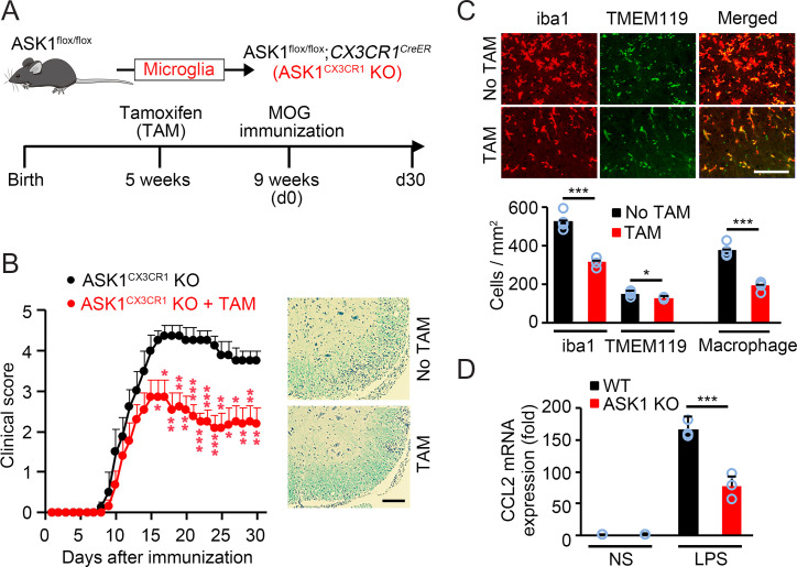
ASK1 deficiency in microglia nearly reproduced the phenotype observed in ASK1LysM KO mice. (A) Schematic diagram illustrating the breeding strategy and experimental design for cell-specific ASK1 knockout in microglia (ASK1CX3CR1 KO). (B) Clinical scores of ASK1CX3CR1 KO EAE mice with or without preadministered TAM (n = 13 and n = 8, respectively). Representative spinal cord sections stained with LFB and H&E are shown at Right. Two-tailed Student’s t test was used. ***P < 0.001; **P < 0.01; *P < 0.05. (Scale bar: 100 µm.) (C) Immunohistochemical staining of iba1 and TMEM119 in the white matter of the spinal cords of ASK1CX3CR1 KO EAE mice killed on their EAE onset day. Quantitative analysis of iba1-positive and TMEM119-positive cells is shown (Lower). Macrophage numbers were deduced from iba1-positive microglia/macrophage and TMEM119-positive microglia numbers. Two-tailed Student’s t test was used. n = 4 mice per group. ***P < 0.001. *P < 0.05. (Scale bar: 100 µm.) (D) mRNA expression of CCL2 in WT and ASK1 KO microglia stimulated with LPS (100 ng/mL) for 6 h. NS: nonstimulated. Experiments were carried out in a 96-well plate format with 3 wells used for each culture condition. Experiments were repeated three times and representative results are shown. Two-tailed Student’s t test was used. ***P < 0.001.

