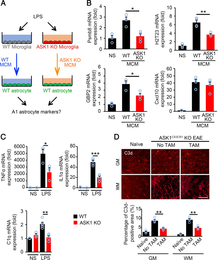
Fig. 4.
ASK1 deficiency in microglia reduced induction of A1 astrocytes in vitro and in vivo. (A) Schematic representation of the experimental procedure for A1 astrocyte induction. (B) A1 astrocyte marker expression (Psmb8, H2T23, and GBP2) in WT astrocytes stimulated with WT MCM or ASK1-deficient MCM for 6 h. The expression of Cxcl10, one of pan-reactive astrocyte markers, was also quantified. Experiments were carried out in a 96-well plate format with 3 to 4 wells used for each culture condition. Experiments were repeated three times and representative results are shown. The one-way ANOVA with Tukey–Kramer post hoc test was used. **P < 0.01; *P < 0.05. (C) TNFα, IL1α, and C1q mRNA expression in WT and ASK1 KO microglia stimulated with LPS (100 ng/mL) for 6 h. NS: nonstimulated. Experiments were carried out in a 96-well plate format with 3 to 4 wells used for each culture condition. Experiments were repeated three times and representative results are shown. Two-tailed Student’s t test was used. ***P < 0.001; **P < 0.01; *P < 0.05. (D) Immunohistochemical staining of C3d in the gray matter (GM) and white matter (WM) of the spinal cords of ASK1CX3CR1 KO EAE mice with or without preadministered TAM on d30 and quantification of the C3d-positive area. The one-way ANOVA with Tukey–Kramer post hoc test was used. n = 4 for WT normal group, n = 6 for ASK1CX3CR1 KO EAE group with or without TAM preadministration. **P < 0.01. (Scale bar: 60 µm.)

