Fig. 5.
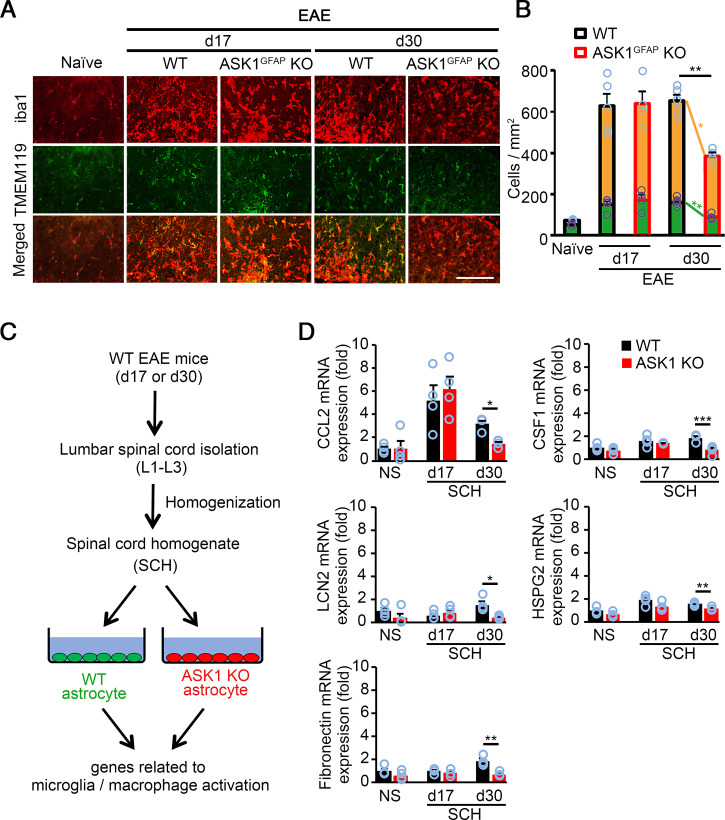
Lack of ASK1 signaling in astrocytes reduces factors that stimulate microglia/macrophage recruitment and activation at d30, but not d17. (A) Immunohistochemical staining of iba1 and TMEM119 in the spinal cords of WT and ASK1GFAP KO EAE mice on d17 and d30. (Scale bar: 100 µm.) (B) Quantification of microglia (green bars) and macrophage (yellow bars) cell numbers in the white matter of the spinal cords. Macrophage numbers were deduced from iba1-positive microglia/macrophage and TMEM119-positive microglia numbers. Two-tailed Student’s t test was used. n = 4 to 5 mice per group. **P < 0.01; *P < 0.05. (C) Schematic representation of the experimental procedure. SCHs taken from WT EAE mice on d17 and d30 were used to stimulate astrocytes. (D) qPCR analysis of CCL2, CSF1, LCN2, HSPG2, and fibronectin in WT and ASK1 KO astrocytes stimulated with 25 µg/mL of SCH for 6 h. Experiments were carried out in a 96-well plate format with 3 to 4 wells used for each culture condition. Experiments were repeated three times and representative results are shown. Two-tailed Student’s t test was used. ***P < 0.001; **P < 0.01; *P < 0.05.

