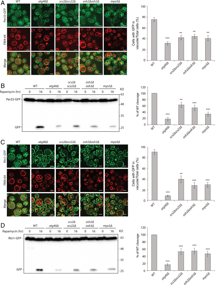
Fig. 2.
Proteins linking the cER to endocytic sites are important for ER-phagy. (A) Scs2/Scs22, Osh2/Osh3, and Myo5 are required for the delivery of Per33 into the vacuole. Left: Fluorescence images of cells expressing Per33-GFP. Cells were treated with rapamycin for 16 h, and vacuoles were stained with fn4-64. Right: The percentage of cells with GFP in the vacuole was quantified. Error bars represent SD, n = 3 independent experiments. **P < 0.01; ***P < 0.001. Student’s t test. (Scale bars, 2 μm.) (B) Left: Western blot of Per33-GFP cleavage after cells were treated with rapamycin for 16 h. Right: Percentage of free GFP divided by the total GFP amount was quantified. WT was set as 100%. All mutants were normalized to the WT control. Error bars represent SD, n = 3 independent experiments. **P < 0.01; ***P < 0.001. Student’s t test. (C) Scs2/Scs22, Osh2/Osh3, and Myo5 are required for the delivery of Rtn1 into the vacuole. Left: Fluorescence images of cells expressing Rtn1-GFP. Cells were treated with rapamycin for 16 h, and vacuoles were stained with fn4-64. Right: The percentage of cells with GFP in the vacuole was quantified. Error bars represent SD, n = 3 independent experiments. **P < 0.01; ***P < 0.001. Student’s t test. (Scale bars, 2 μm.) (D) Left: Western blot analysis of Rtn1-GFP cleavage after cells were treated with rapamycin for 16 h. Right: Percentage of free GFP divided by the total GFP amount was quantified. WT was set as 100%. All mutants were normalized to the WT control. Error bars represent SD, n = 3 independent experiments. ***P < 0.001. Student’s t test.

