Fig. 4.
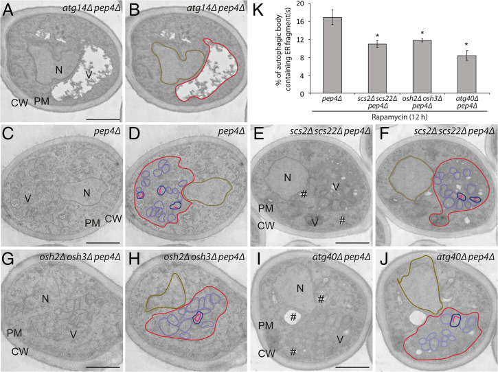
Scs2/Scs22 and Osh2/Osh3 are required for the sequestration of ER fragments into autophagic bodies (ABs). Cells were treated with rapamycin for 12 h and then fixed with permanganate, embedded in Spurr’s resin, and processed for electron microscopy. (A and B) Images of the atg14Δ pep4Δ strain. (C and D) Images of the pep4Δ strain. (E and F) Images of the scs2Δ scs22Δ pep4Δ cells. (G and H) Images of the osh2Δ osh3Δ pep4Δ strain. (I and J) Images of the atg40Δ pep4Δ cells. The location of vacuole (V), nucleus (N), cell wall (CW), PM, and lipid droplet (#) are marked in A, C, E, G, and I. Vacuole (red), nucleus (brown), AB (purple), AB containing ER fragments (blue), and ER fragments inside AB (pink) are outlined in B, D, F, H, and J. (K) Quantitation of the percentage of ABs containing ER. *P < 0.05. Student’s t test. (Scale bars, 1 µm.)

