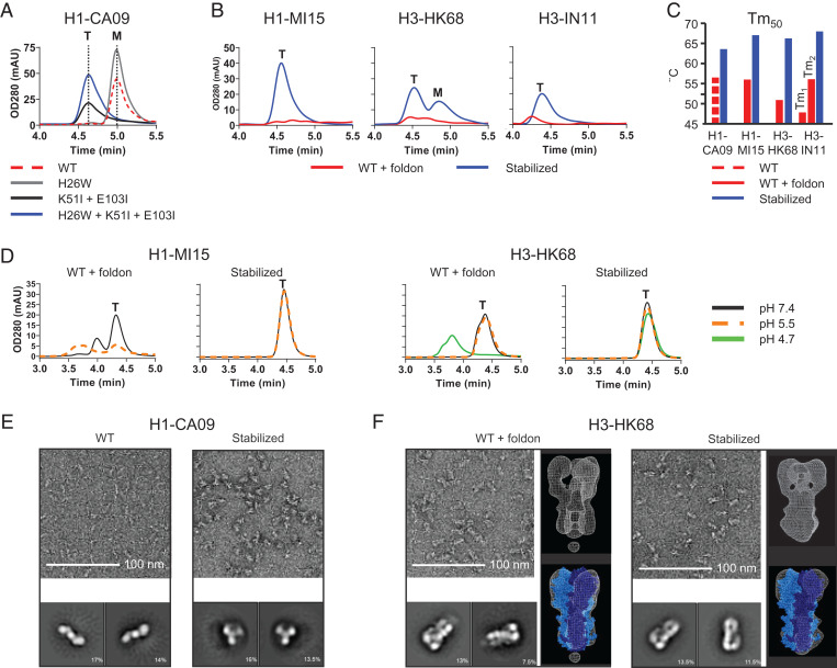
Fig. 2.
Characterization of HA-containing stabilizing mutations. (A) Analytical SEC profiles of cell culture supernatant of Expi293F cells expressing H1-CA09 HA with combinations of stabilizing mutations; WT, H26W substitution, K51I + E103I substitution, and fully stabilized H26W, K51I + E103I. Data are representing mean of three independent transfections in one experiment. Peaks representing monomeric and trimeric species are indicated by an “M” and “T,” respectively. (B) Analytical SEC culture supernatant profiles of Expi293F cells expressing WT HA including foldon trimerization domain and fully stabilized HA (H26W, K15I, and E103I) for H1-MI15, H3-HK68, and H3-IN11. (C) Temperature stability of purified HA determined by DSF. Shown are Tm50 values for WT H1-CA09 and stabilized H1-CA09 HA and for WT foldon-fused and stabilized HA of H1-MI15, H3-HK68, and H3-IN11. (D) Two-week pH stability at 4 °C of purified HA analyzed by analytical SEC. Shown are profiles of WT and stabilized HA of H1-MI15 (pH 5.5 and 7.4) and H3-HK68 (pH 4.7, 5.5, and 7.4). (E) Negative-stained EM of WT H1-CA09 (Left) and stabilized HA (Right) with representative 2D-averaged classes. (F) Negative-stained EM of WT H3-HK68 HA foldon-fused (Left) and stabilized HA (Right) with representative 2D-averaged classes (each box is 27.4 nm) and 3D density map (white mesh) superimposed on the X-ray structure of H3-HK68 HA trimer (PDB identifier 4FNK) represented in blue spheres with one monomer plotted in dark blue.

