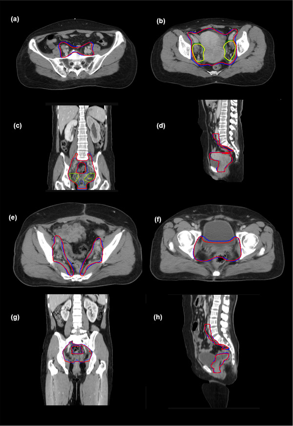
FIGURE 3.

Comparison of the results between automatic segmentations and reference contours. (a and b) clinical tumor volume for definitive radiotherapy (dCTV)1 and dCTV2 in different cross‐sections, (c) coronal view, and (d) sagittal view. dCTV1 and dCTV2 of the reference are in red and yellow, respectively. dCTV1 and dCTV2 of the automatic segmentation are in blue and green, respectively; (e and f) clinical tumor volume for postoperative radiotherapy (pCTV)1 in different cross‐sections, (g) coronal view, and (h) sagittal view. pCTV1s of the reference and automatic segmentation are in red and blue, respectively
