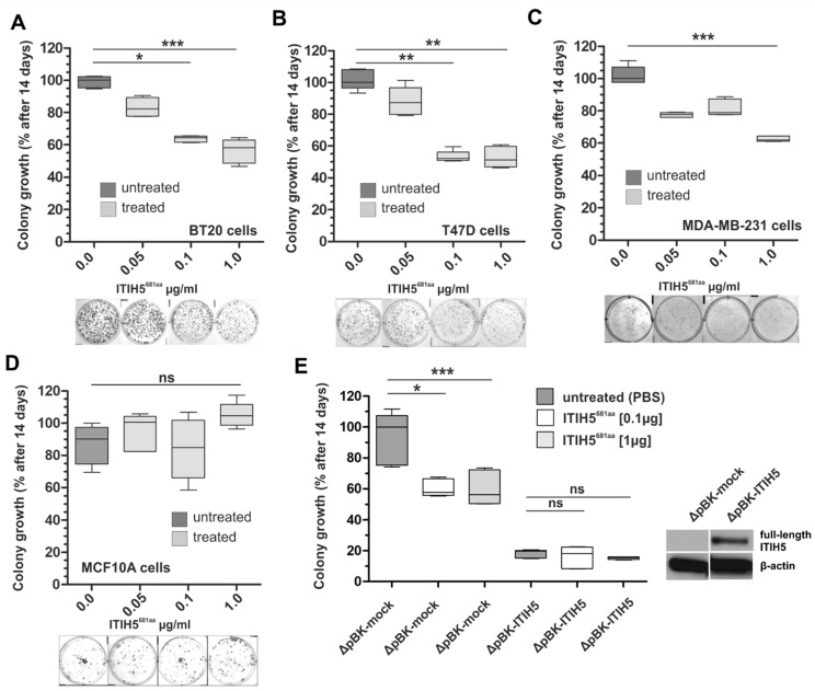
Figure 3.
N-terminal ITIH5t protein (ITIH5681aa) suppressed the colony growth of malignant breast cancer cell lines but not of the benign breast cell line MFC10A. (A–D) A clonogenic survival assay was performed under N-terminal ITIH5t application over 14 days. The dose-dependent suppressive effect of N-terminal ITIH5t on colony formation is shown as a box plot and by representative growth colonies (below graphs) for (A) basal BT20 (B) luminal T47D and (C) basal MDA-MB-231 breast cancer cells. (D) The suppressive impact of N-terminal ITIH5t on benign MCF10A breast cancer cells was not observed. * p ≤ 0.05; ** p ≤ 0.01; *** p ≤ 0.001; ns: not significant (p > 0.05). (E) Based on in vitro models of breast cancer cells stably re-expressing ITIH5 (ΔpBK-ITIH5) and the corresponding mock clones (ΔpBK-mock) (see also Figure 1 and Rose et al. [7]), clonogenic survival assays were performed under N-terminal ITIH5t application (0.1 and 1.0 µg/mL) to assess the specificity. Only mock control clones lacking ITIH5 expression were significantly responsible for the suppressive effects mediated by the applied N-terminal ITIH5t. ΔpBK-ITIH5 clones overexpressing the full-length ITIH5 protein were not further affected by the N-terminal ITIH5t treatment. Note that growth differences between the mock clones (ΔpBK-mock) treated with ITIH5t and the overexpressing clones (ΔpBK-ITIH5) could be caused by various variables, including the different exposure times of the administered vs. endogenously expressed proteins.

