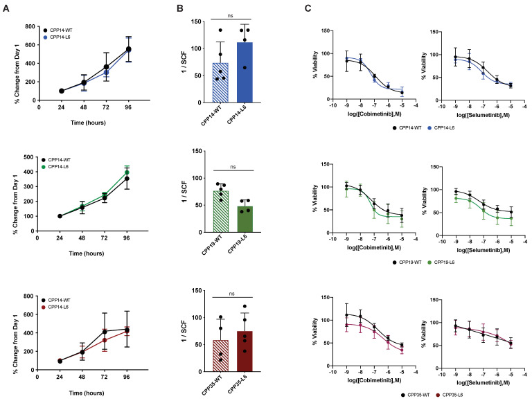
Figure 2.
Optical barcoding does not perturb the biological properties of wild-type cells. Three patient-derived cell lines (CPP14, blue; CPP19, green; and CPP35, red) were optically barcoded (denoted by “L6” variants). (A) Proliferation was assessed via a resazurin metabolic assay performed at 24 h intervals for a period of 96 h (n = 4 independent experiments, mean ± SD). Doubling time (hours) ± 95% CI for the WT and L6 pairs were calculated by fitting an exponential growth equation (ns = non-significant, see Table S1). (B) ELDA performed to determine the SCF of the WT vs L6 cell pairs (n = 4 independent experiments, mean ± SD). The presence or absence of colonospheres was assessed 10 days after seeding at densities of 1000, 1000, 10 or 1 cells per well and is reported as the mean SCF ± SD (P = ns, Student’s t-test). (C) WT and L6 cells were treated with escalating doses of the MEK inhibitors cobimetinib and Selumetinib for 72 h, at which point cell viability was assessed via resazurin assay. Data is normalized to the vehicle control for each compound and reported as mean % viability ± SD (n > 4). LogIC50 ± 95% CI values were calculated by interpolating sigmoidal dose-response curves (see Table S2). WT, wild-type; ELDA, Extreme limiting dilution analysis; SCF, stem cell frequency.

