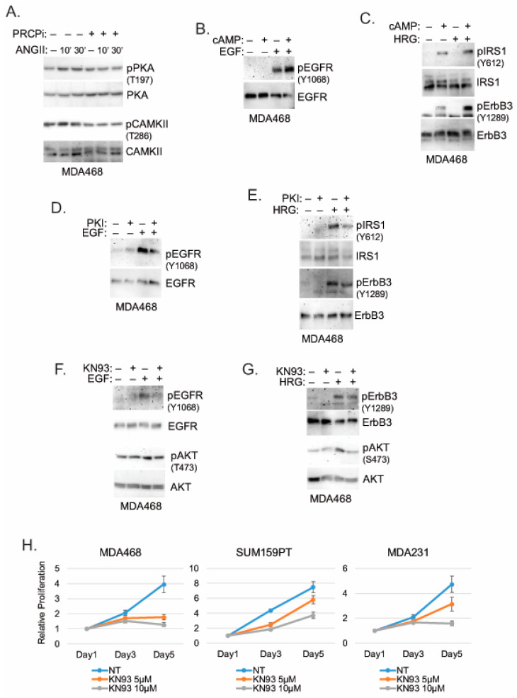
Figure 7.
PRCPi suppresses PKA and CAMKII activation to decrease activation of EGFR/ErbB3 and IRS1. (A) MDA468 cells were serum-starved and pretreated with a vehicle or PRCPi (10 µM) and then treated with Ang II (100 nM) for the indicated times. Serum-starved MDA468 cells were treated with cAMP and/or EGF (B), cAMP and/or HRG (C), PKI and/or EGF (D), PKI and/or HRG (E), KN93 and/or EGF (F), or KN93 and/or HRG (G) for 10 min. Lysates were immunoblotted for the indicated proteins. (H) The indicated cell lines were cultured in the presence of the vehicle or KN93 (5 µM and 10 µM) and were harvested at the indicated times. Relative cell proliferation was analyzed with Hoechst staining of cellular DNA. Average (8 replicate) relative fluorescence intensity of Hoechst is presented with SD indicated. There is significant differences (p = 0.000) between the vehicle and 10 µM KN93 conditions in all cell lines. There is a significant difference (p = 0.000) between the vehicle and 5 µM KN93 in MDA468 cells. There are no significant differences (p > 0.1) between the vehicle and 5 µM KN93 conditions in SUM159PT and MDA231 cells. Original Western Blots and densitometry can be found at Figures S12–S14.

