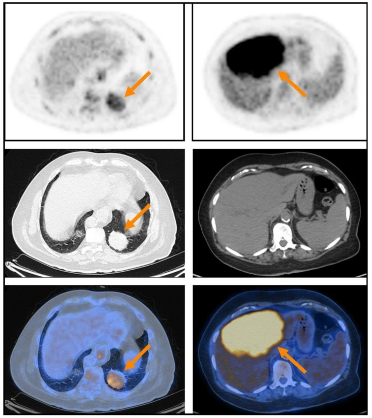
Figure 1.
Two illustrative extranodal MALT lymphoma lesions that were detected on PET (upper row), presented together with corresponding CT (middle row) and fusion PET-CT (lower row) transaxial slices: a case of lung MALT lymphoma with lower [18F]FDG-avidity (SUVmax 5.9) on the left, and a case of liver MALT lymphoma with higher [18F]FDG-avidity (SUVmax 9.0) on the right. Both patients were referred to systemic therapy after imaging. While the patient with the lung lesion did not progress during a follow-up period of 6.4 years, the patient with the liver lesion progressed 2.4 years after staging.

