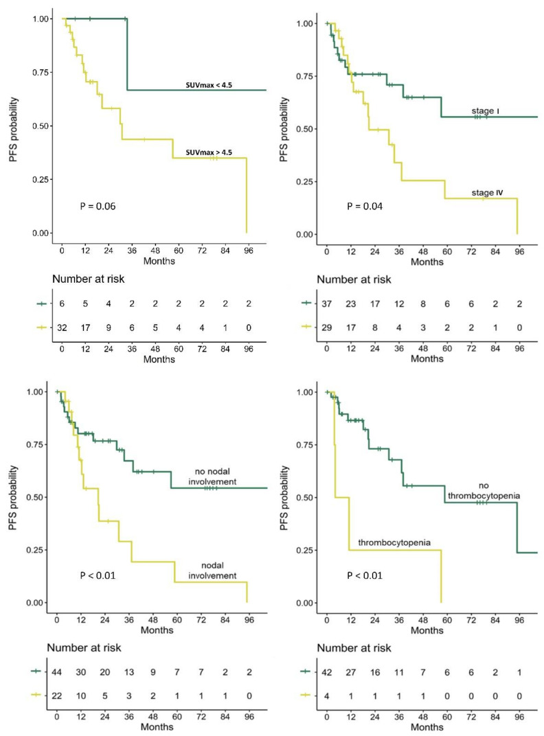
Figure 2.
Kaplan–Meier curves of PFS probability in (upper left) patients whose extranodal MALT lymphoma lesions were detected on PET and had SUVmax greater vs lower than 4.5, (upper right) patients with stage I vs IV disease, (lower left) patients with vs without nodal involvement, and (lower right) patients with vs without thrombocytopenia. PFS, progression-free survival; SUVmax, maximum standardized uptake value; Log-rank p-value is presented for each survival analysis.

