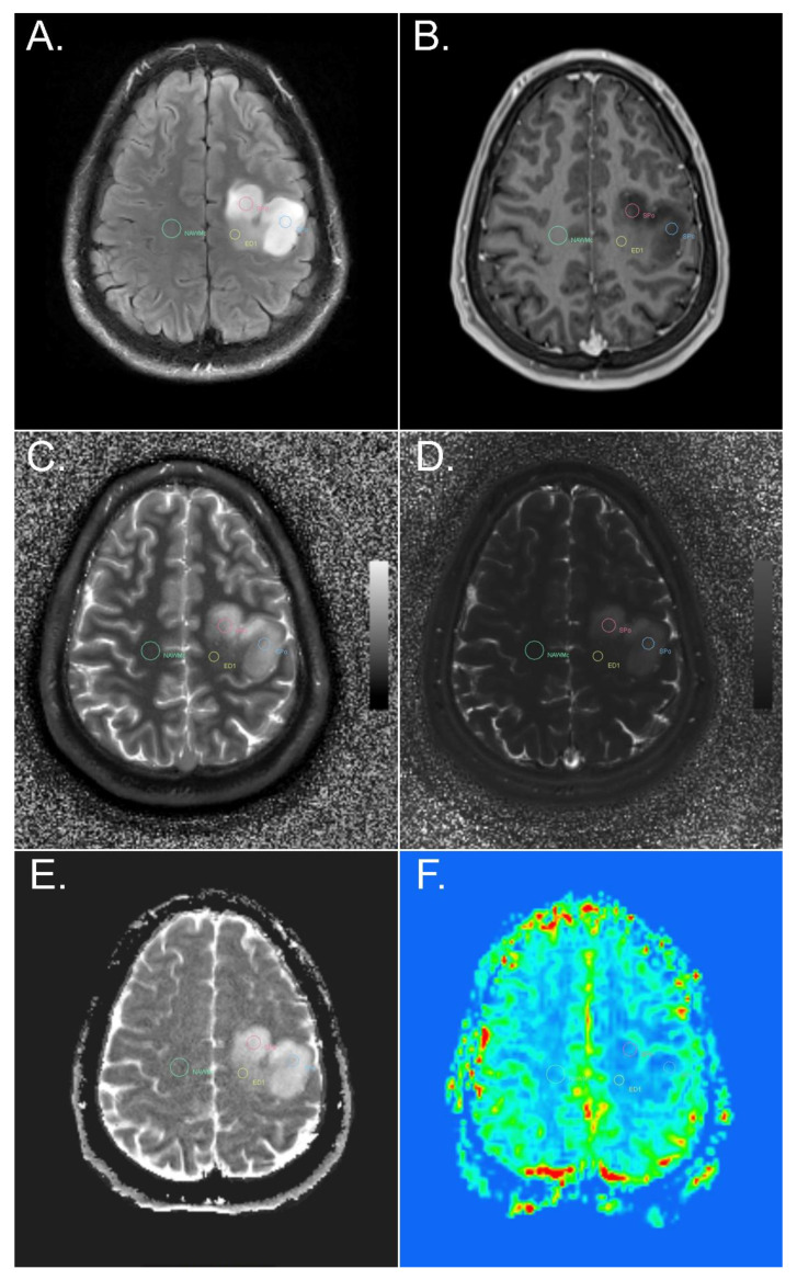
Figure 1.
Example of ROI placement in different tumor and peritumoral components of a diffuse astrocytoma, IDH-mutant, in a left precentral location. FLAIR (A), T1-weighted MPR post i.v. contrast administration (B), MRF T1 (C) and T2 (D) maps, ADC map (E), and perfusion-weighted imaging (F). ROIs were placed in the solid tumor part without contrast enhancement and without an increase in rCBV (red and blue circle), in peritumoral edema (yellow circle), and in contralateral NAWM (green circle). SPo, solid part of the tumor without contrast enhancement; ED1, peritumoral edema less than or equal to 1 cm distant from the tumor; NAWMc, contralateral NAWM of the frontal lobe.

