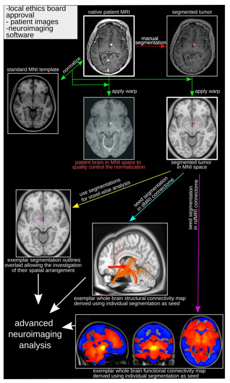
Figure 2.
Framework for normative brain analyses. Following typical research project prerequisites (upper left side of the image), the analysis begins with the native patient MRI. The feature of interest (e.g., tumor) is manually segmented (red arrow) using the native patient image. The native patient brain is then normalized (transformed) to MNI space and the estimated transforms applied to the native patient brain (for quality control) and the segmented feature (green arrows). The segmented feature (e.g., tumor) in MNI space is the main input for further processing, such as voxel-based group analysis (yellow arrow), and is used as seeds in normative structural (turquoise arrow) and functional (purple arrow) connectome analyses to derive brain-wide connectivity patterns. dMRI = diffusion-weighted MRI; MNI = Montreal Neurological Institute; MRI = Magnetic resonance imaging; rsfMRI = resting state functional MRI.

