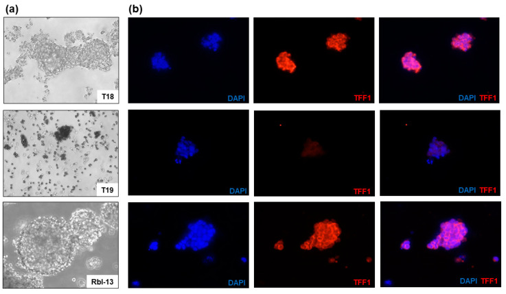
Figure 3.
Immunofluorescence TFF1 staining of two primary RB cell cultures (T18 and T19) and RB cell line Rbl-13. (a) Morphology of the primary RB cell cultures T18 and T19 as well as RB cell line Rbl-13 revealed by phase contrast imaging (200×). (b) DAPI (blue), TFF1 (red), and merged DAPI/TFF1 immunofluorescence staining of the respective RB cells (200×). T18 and the positive control Rbl-13 showed a high expression of TFF1 in contrast to T19 cells without TFF1 expression.

