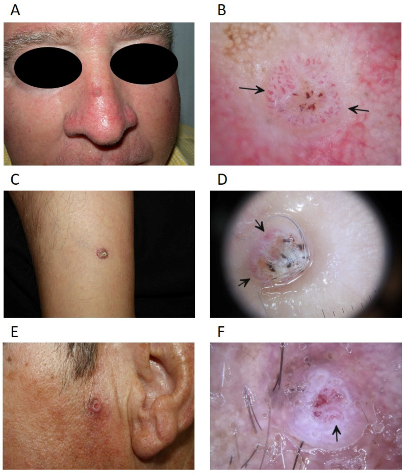
Figure 2.
Dermoscopy of cutaneous squamous cell carcinoma. (A) Wart-like tumour lesion on the dorsum of the nose; (B) dermoscopy with polarised light showing a predominantly vascular pattern with serpentine, hairpin and irregular vessels (arrows), central ulceration and blood staining; (C) crateriform keratinising tumour lesion; (D) polarised light dermoscopy with central whitish crust and presence of irregular and comma-shaped vessels (arrows) in the periphery; (E) crateriform tumour lesion; (F) polarised light dermoscopy showing white unstructured areas with irregular groups of white perifollicular circles, central vascular pattern with hairpin and irregular vessels (arrows).

