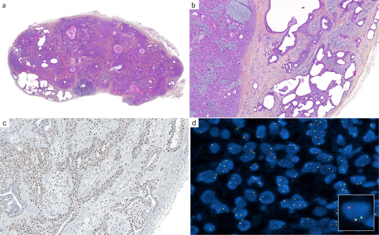
Figure 2.
Cutaneous mixed tumor (chondroid syringoma): (a) cutaneous mixed tumor presenting as a circumscribed nodule, with cysts, ducts, nodules, and abundant stroma (×25); (b) higher magnification reveals different cell populations: myoepithelial aggregates (left), elaborated and cystic ductal structures (right), and mesenchymal cells associated with fibrous, fibromyxoid, and myxoid stroma (×100); (c) PLAG1 immunohistochemistry: diffuse nuclear staining is present, notably in the myoepithelial component (×200); (d) fluorescence in situ hybridization (FISH, break-apart probe) demonstrates a clonal PLAG1 gene rearrangement (×1000).

